#ionized injection system
Explore tagged Tumblr posts
Text
Into Jag Crag Gorge

STAR WARS EPISODE I: The Phantom Menace 01:01:32
#Star Wars#Episode I#The Phantom Menace#Tatooine#Boonta Eve Classic#podrace#Jag Crag Gorge#Gasgano's podracer#Sebulba's podracer#quold runium#Ord Pedrovia#Plug-F Mammoth#ionized injection system#Split-X repulsor generator housing#Split-X stabilizing vane#Teemto Pagalies' podracer#IPG-X1131 LongTail
1 note
·
View note
Text

Was the additive A-50 used by the CIA and the Air Force on their Blackbirds?
Dr. Richard Bissell, the CIA’s Special Assistant for Planning and Coordination, who was managing the program, was so worried about this particular issue, (hiding the plume of the afterburner) that he had considered calling for the scrapping of the entire development of a U-2 successor. ( it was a high priority for the CIA to disguise the A-12 as much as possible. The A-50 additive option is what pushed the CIA into giving the contract to Lockheed instead of Convair) Linda Sheffield
That’s where the cesium additive, which eventually became known as A-50, came in an idea that Lovick claims saved the A-12 program.
The basic principle behind this is a concept known as “plasma stealth.” In the simplest terms, this involves creating a cloud of plasma, or ionized gas, around some or all of an object. The plasma then absorbs electromagnetic radiation, such as radar waves, preventing them from reflecting. There are multiple ways to generate the required plasma Lovick’s idea was to inject an alkali metal, via a fuel additive, into the extremely hot exhaust streams, where the heat would turn it into an ionized gas. The question remains today did they use it or not? There is evidence pointing in both directions. Also, was this additive used in the SR 71?
I think this is highly possible as they converted KC 135s to be able to carry the additive with some degree of safety. The segregated fuel system on the KC-135Q tankers, which the Air Force eventually re-engined with ( A-50 ) CFM56s to create the KC-135T variant, would have largely eliminated the concerns about the additive finding its way into the tanker’s engines for any reason. KC-10 Extenders, which also supported the Blackbirds later in their service life, also could keep the JP-7 separated from the aircraft’s fuel load.
My father, Butch Sheffield a Pioneer in the SR 71 program in his notes. Discussed the additive and admitted that it could’ve been toxic to humans.
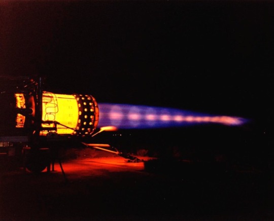
This picture shows an A-12 flying over Area 51 in Nevada or nearby that area. What is being emitted from the back of the A-12 is a combination of JP-7 fuel and heat exhaust it was not just dumping fuel. The other photo is a picture of the J 58 engine in a test mode which shows how much energy and power was emitted from the engines of the A-12 and the SR -71 that were hard to disguise but was possible with the additive A-50.
If nothing else, though, the development of the A-50 additive is another intriguing and very obscure part of the story of the A-12, as well as its successor, the SR-71, much of which remains unknown to this day, and it also offers intriguing insights into the dawn of stealth technology.
@Habubrats71 via X
#sr 71#sr71#sr 71 blackbird#blackbird#aircraft#usaf#lockheed aviation#skunkworks#aviation#mach3+#habu#reconnaissance#cold war aircraft
50 notes
·
View notes
Text


Mining old data from NASA's Voyager 2 solves several Uranus mysteries
When NASA's Voyager 2 spacecraft flew by Uranus in 1986, it provided scientists' first—and, so far, only—close glimpse of this strange, sideways-rotating outer planet. Alongside the discovery of new moons and rings, baffling new mysteries confronted scientists. The energized particles around the planet defied their understanding of how magnetic fields work to trap particle radiation, and Uranus earned a reputation as an outlier in our solar system.
Now, new research analyzing the data collected during that flyby 38 years ago has found that the source of that particular mystery is a cosmic coincidence. It turns out that in the days just before Voyager 2's flyby, the planet had been affected by an unusual kind of space weather that squashed the planet's magnetic field, dramatically compressing Uranus's magnetosphere.
"If Voyager 2 had arrived just a few days earlier, it would have observed a completely different magnetosphere at Uranus," said Jamie Jasinski of NASA's Jet Propulsion Laboratory in Southern California and lead author of the new work published in Nature Astronomy. "The spacecraft saw Uranus in conditions that only occur about 4% of the time."
Magnetospheres serve as protective bubbles around planets (including Earth) with magnetic cores and magnetic fields, shielding them from jets of ionized gas—or plasma—that stream out from the sun in the solar wind. Learning more about how magnetospheres work is important for understanding our own planet, as well as those in seldom-visited corners of our solar system and beyond.
That's why scientists were eager to study Uranus's magnetosphere, and what they saw in the Voyager 2 data in 1986 flummoxed them. Inside the planet's magnetosphere were electron radiation belts with an intensity second only to Jupiter's notoriously brutal radiation belts. But there was apparently no source of energized particles to feed those active belts; in fact, the rest of Uranus's magnetosphere was almost devoid of plasma.
The missing plasma also puzzled scientists because they knew that the five major Uranian moons in the magnetic bubble should have produced water ions, as icy moons around other outer planets do. They concluded that the moons must be inert with no ongoing activity.
Solving the mystery
So why was no plasma observed, and what was happening to beef up the radiation belts? The new data analysis points to the solar wind. When plasma from the sun pounded and compressed the magnetosphere, it likely drove plasma out of the system. The solar wind event also would have briefly intensified the dynamics of the magnetosphere, which would have fed the belts by injecting electrons into them.
The findings could be good news for those five major moons of Uranus: Some of them might be geologically active after all. With an explanation for the temporarily missing plasma, researchers say it's plausible that the moons actually may have been spewing ions into the surrounding bubble all along.
Planetary scientists are focusing on bolstering their knowledge about the mysterious Uranus system, which the National Academies' 2023 Planetary Science and Astrobiology Decadal Survey prioritized as a target for a future NASA mission.
JPL's Linda Spilker was among the Voyager 2 mission scientists glued to the images and other data that flowed in during the Uranus flyby in 1986. She remembers the anticipation and excitement of the event, which changed how scientists thought about the Uranian system.
"The flyby was packed with surprises, and we were searching for an explanation of its unusual behavior. The magnetosphere Voyager 2 measured was only a snapshot in time," said Spilker, who has returned to the iconic mission to lead its science team as project scientist. "This new work explains some of the apparent contradictions, and it will change our view of Uranus once again."
Voyager 2, now in interstellar space, is almost 13 billion miles (21 billion kilometers) from Earth.
TOP IMAGE: NASA’s Voyager 2 captured this image of Uranus while flying by the ice giant in 1986. New research using data from the mission shows a solar wind event took place during the flyby, leading to a mystery about the planet’s magnetosphere that now may be solved. Credit: NASA/JPL-Caltech
LOWER IMAGE: The first panel of this artist’s concept depicts how Uranus’s magnetosphere — its protective bubble — was behaving before the flyby of NASA’s Voyager 2. The second panel shows an unusual kind of solar weather was happening during the 1986 flyby, giving scientists a skewed view of the magnetosphere. Credit: NASA/JPL-Caltech
7 notes
·
View notes
Text
Ultrasound Test Cost in Delhi NCR
Ultrasound, also known as sonography, is a widely used imaging technique that plays a crucial role in modern medical diagnostics. By using high-frequency sound waves to produce real-time images of the inside of the body, ultrasound offers a safe, painless, and non-invasive way to evaluate organs, tissues, and blood flow. It is used in a wide range of medical specialties, from obstetrics to cardiology, and continues to be one of the most accessible and cost-effective diagnostic tools available today.
1. Safe and Non-Invasive Procedure
One of the most important benefits of ultrasound is its safety. Unlike imaging techniques that use ionizing radiation, such as X-rays or CT scans, ultrasound relies solely on sound waves. This makes it completely radiation-free and safe for repeated use. It is especially beneficial for sensitive patient groups like pregnant women, unborn babies, and young children.
Ultrasound is also non-invasive, which means there is no need for incisions or injections in most cases. This reduces patient discomfort and eliminates the risk of infection associated with invasive procedures.
2. Widely Used in Pregnancy Monitoring
Ultrasound is best known for its critical role in obstetrics and gynecology. During pregnancy, it is used to monitor the development and health of the fetus. Doctors can check the baby's growth, heartbeat, position, and even detect congenital anomalies early. Parents often look forward to ultrasound scans as they provide the first images of their unborn child.
Ultrasound can also help estimate the due date, determine the number of fetuses, check for ectopic pregnancy, and monitor amniotic fluid levels.
3. Real-Time Imaging
Ultrasound provides real-time images, which is incredibly useful for both diagnosis and guidance during certain procedures. This dynamic imaging allows healthcare providers to observe the movement of internal organs, heartbeats, and blood flow in real-time. It’s also used to guide procedures such as biopsies, fluid drainage, and catheter placement, ensuring accuracy and reducing complications.
4. Versatile Diagnostic Applications
Ultrasound is a versatile tool that can be used to examine various parts of the body, including:
Abdomen: Liver, gallbladder, kidneys, pancreas, and spleen.
Pelvis: Uterus, ovaries, bladder, and prostate.
Heart: Echocardiograms assess heart function and detect heart conditions.
Musculoskeletal System: Muscles, tendons, and joints for injuries or inflammation.
Blood Vessels: Doppler ultrasound checks for blood clots, blockages, or poor circulation.
Its wide range of applications makes ultrasound an indispensable part of diagnostic medicine.
5. Quick and Accessible
Ultrasound exams are generally quick, often completed in 15–30 minutes. Results are usually available promptly, which helps doctors make immediate decisions in emergency situations. Additionally, ultrasound machines are portable and can be used at the bedside in intensive care units or even in remote areas, making healthcare more accessible.
6. Cost-Effective Imaging Option
Compared to other imaging technologies such as MRI and CT scans, ultrasound is far more affordable. This makes it ideal for routine screening and widespread use, especially in developing countries where advanced imaging technologies may not be readily available. It provides reliable diagnostic information at a fraction of the cost, benefiting both healthcare providers and patients.
7. No Known Side Effects
Ultrasound is considered extremely safe and has no known long-term side effects. It doesn’t expose the patient to any harmful elements and is well-tolerated across all age groups. This safety profile makes it ideal for repeated monitoring in chronic conditions or pregnancy.
8. Helpful in Preventive Healthcare
Ultrasound can be a valuable tool in preventive healthcare. Regular abdominal or pelvic ultrasound scans can detect early signs of diseases such as liver cirrhosis, kidney stones, ovarian cysts, or fibroids. Early detection leads to early treatment, improving patient outcomes and preventing complications.
Ultrasound stands out as a safe, effective, and versatile imaging tool in modern healthcare. Its non-invasive nature, affordability, and broad range of applications make it one of the most commonly used diagnostic methods worldwide. Whether it’s monitoring a growing fetus, diagnosing organ conditions, or guiding critical procedures, ultrasound continues to play a key role in accurate and timely diagnosis.
For patients, ultrasound offers peace of mind—quick results without discomfort or risk. For doctors, it provides essential insights that guide treatment decisions. As technology advances, ultrasound continues to evolve, offering even more precise imaging and expanding its impact across all areas of medicine.
To find out the ultrasound test cost in Delhi visit Healthi India
0 notes
Text
Lipid Contract Manufacturing Market Growth Analysis, Market Dynamics, Key Players and Innovations, Outlook and Forecast 2025-2032
According to a new report from Intel Market Research, the global lipid contract manufacturing market was valued at USD 753 million in 2024 and is projected to reach USD 699 million by 2032, exhibiting a negative CAGR of 1.1% during the forecast period (2025–2032). This market correction reflects supply chain normalization post-COVID vaccine boom, yet retains strong fundamentals in specialized drug delivery applications.
📥 Download FREE Sample Report: Lipid Contract Manufacturing Market - View in Detailed Research Report
What is Lipid Contract Manufacturing?
Lipid contract manufacturing��involves the outsourced production of advanced drug delivery systems utilizing lipid nanoparticles (LNPs), liposomes, and other lipid-based formulations. These technologies serve as protective carriers for therapeutics, particularly benefiting fragile payloads like mRNA, siRNA, and gene therapies. The field has gained prominence through critical applications in COVID-19 vaccines, where LNPs proved indispensable for mRNA delivery.
The manufacturing process requires specialized expertise in:
Precision formulation of ionizable lipids, phospholipids, and cholesterol blends
Microfluidic production technologies for consistent particle sizing
GMP-compliant aseptic processing for sterile injectables
Leading CDMOs have established themselves as essential partners for biopharma companies developing next-generation therapies, particularly in oncology, rare diseases, and genetic medicines.
Key Market Drivers
1. Accelerated Adoption of mRNA Therapeutics Beyond Vaccines
The successful deployment of LNP-formulated COVID vaccines—over 10 billion doses administered globally—validated the platform's potential. Pharmaceutical companies are now actively developing mRNA applications for:
Oncology: Personalized cancer vaccines like Moderna's mRNA-4157 combined with Keytruda®
Rare diseases: Protein replacement therapies for metabolic disorders
Infectious diseases: Improved flu vaccines and emerging pathogen readiness
This therapeutic expansion drives sustained demand for specialized lipid manufacturing capabilities, particularly for novel ionizable lipids that improve delivery efficiency and reduce side effects.
2. Biopharma's Strategic Shift Toward Outsourcing
Pharmaceutical companies now allocate 25-30% of manufacturing budgets to external partners, with lipid nanoparticle production being one of the most frequently outsocused functions. This trend stems from:
The $50-75 million capital cost for in-house LNP facilities
Shortage of lipid formulation experts—currently less than 300 globally with deep LNP experience
Regulatory advantages of partnering with CDMOs holding existing FDA/EMA approvals
Recent deals like Merck's $780 million acquisition of Exelead underscore the strategic value placed on lipid manufacturing expertise.
Market Challenges
The sector faces several headwinds requiring careful navigation:
1. Regulatory Complexity for Novel Lipids
Each new lipid excipient requires full safety characterization—a process taking 18-24 months longer than conventional drug formulations. The FDA's 2023 draft guidance on lipid nanoparticles establishes rigorous standards for:
Impurity profiling to 0.1% levels
Long-term stability under various stress conditions
Comprehensive immunogenicity assessments
2. Supply Chain Vulnerabilities
Critical lipids like DMG-PEG2000 remain dependent on just 2-3 global suppliers, creating risks that manufacturers mitigate through:
Strategic stockpiling (6-12 month inventories)
Dual-sourcing arrangements
In-house synthesis capabilities for key components
Emerging Opportunities
Innovation is unlocking new frontiers beyond vaccines:
1. Targeted Delivery Breakthroughs
Next-generation LNPs with tissue-specific targeting ligands (e.g., galactose for liver delivery) enable:
Reduced systemic toxicity for chemotherapeutics
Improved biodistribution for gene editing tools
Enhanced CNS penetration for neurological therapies
2. Asia-Pacific Market Expansion
Countries like China and South Korea are making strategic investments:
$2.1 billion committed to biomanufacturing infrastructure in China's 14th Five-Year Plan
South Korea's Samsung Biologics expanding into lipid formulations
India emerging as cost-competitive alternative for preclinical-stage production
📘 Get Full Report Here: Lipid Contract Manufacturing Market - View in Detailed Research Report
Competitive Landscape
The market features specialized players across the value chain:CompanyKey StrengthRecent DevelopmentMerck KGaAEnd-to-end mRNA solutionsExelead integration completed 2023Precision NanoSystemsMicrofluidic platform technologyLaunched GMP NanoAssemblr® system in 2024EvonikSustainable lipid production€150M German facility expansionCordenPharmaCold chain expertiseNew -80°C storage capabilities
Smaller innovators like Acuitas Therapeutics and Genevant Sciences compete through proprietary lipid libraries and targeted delivery solutions.
Technology Frontiers
Innovation focuses on overcoming key limitations:
1. Scalability Solutions
Traditional microfluidics face challenges in commercial-scale production. New approaches include:
Continuous flow manufacturing systems (e.g., Precision NanoSystems' NxGen™)
Adaptive mixing technologies allowing 10-100x scale-up from clinical batches
2. Lyophilization Advances
Enabling room-temperature stability through:
Novel cryoprotectant formulations
Controlled nucleation during freezing
Optimized reconstitution protocols
Recent publications demonstrate successful lyophilization of LNP-formulated mRNA with ≥90% recovery of biological activity.
Regional Insights
North America dominates (60% share) through:
Concentration of mRNA technology pioneers (Moderna, Pfizer)
FDA's progressive stance on novel delivery systems
Strong venture funding for lipid-focused biotechs
Europe maintains strength with:https://www.intelmarketresearch.com/life-sciences/712/global-premenstrual-syndrome-treatment-forecast-2025-2032
Merck's integrated capabilities in Darmstadt
Academic excellence at ETH Zurich and KU Leuven
EMA's centralized approval pathway
Visit more reports :
About Intel Market Research
Intel Market Research is a leading provider of strategic intelligence, offering actionable insights in biotechnology, pharmaceuticals, and healthcare infrastructure. Our research capabilities include:
Real-time competitive benchmarking
Global clinical trial pipeline monitoring
Country-specific regulatory and pricing analysis
Over 500+ healthcare reports annually
Trusted by Fortune 500 companies, our insights empower decision-makers to drive innovation with confidence.
🌐 Website: https://www.intelmarketresearch.com 📞 International: +1 (332) 2424 294 📞 Asia-Pacific: +91 9169164321 🔗 LinkedIn: Follow Us
0 notes
Text
HPLC Columns
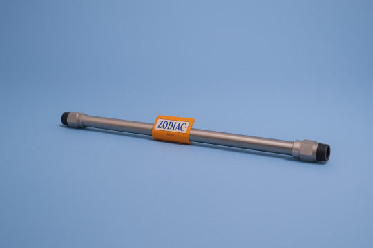
High-performance liquid chromatography (HPLC) is a crucial technique in modern analytical chemistry. The column, a complex part of every HPLC system, plays a vital role in separating intricate chemical mixtures.
An HPLC column functions like a highly selective filter. As your sample passes through the column, various compounds separate according to their distinct chemical characteristics. This separation occurs through specific interactions between the components of your sample and the specialized material packed within the column.
In this comprehensive guide, you'll discover:
The essential components and structure of HPLC columns
Different types of columns and their specific applications
Key factors in selecting the right column for your analysis
Practical tips for maintaining column performance
Expert answers to common HPLC column questions
Whether you're new to chromatography or aiming to enhance your current methods, understanding HPLC columns is vital for obtaining precise and dependable results in your analytical work. Let's delve into the captivating realm of HPLC columns and unleash their complete potential in your laboratory applications.
Understanding HPLC Columns: Structure and Function
HPLC columns feature a robust stainless steel housing designed to withstand high pressures during analysis. Inside this metallic shell lies the heart of the column - a densely packed bed of particles known as the stationary phase.
Structure of HPLC Columns
HPLC columns are made up of two main components:
Stainless Steel Housing: The outer shell of the column, which is designed to withstand high pressures during analysis.
Stationary Phase: The inner bed of particles that is responsible for separating the different components of a sample.
The stationary phase consists of specialized particles, typically made from porous silica. These particles measure between 1.7 and 5 micrometers in diameter and possess a unique characteristic: their surface contains countless microscopic pores. This porous structure creates an immense surface area - a single gram of these particles can provide up to 400 square meters of surface area.
How HPLC Columns Work
The mobile phase - a liquid solvent - flows through the spaces between these packed particles. As your sample travels through the column, different compounds interact with the stationary phase's surface chemistry in unique ways. These interactions occur across the vast surface area provided by the porous particles, creating effective separation conditions.
The combination of precise particle packing, extensive surface area, and specific surface chemistry enables HPLC columns to achieve high-resolution separation of complex sample mixtures.
How HPLC Columns Work
HPLC columns work by creating a dynamic interaction between the sample mixture and the internal environment of the column. When you inject your sample into the HPLC system, the mobile phase carries it into the column under high pressure.
The separation occurs through two main processes:
1. Partitioning Process
Sample components distribute between the mobile phase and stationary phase
Each compound moves at its unique speed based on chemical properties
Stronger attraction to stationary phase = slower movement
Weaker attraction = faster movement through the column
2. Retention Behavior
Polar compounds stick longer to polar stationary phases
Non-polar molecules travel faster through polar environments
pH affects ionization state and retention patterns
Temperature influences interaction strength
As your sample moves through the column, its components separate at different speeds. You can think of it as a race where each molecule has its own pace. Fast-moving compounds with minimal attraction to the stationary phase exit first, creating early peaks in your chromatogram. Compounds with stronger attractions emerge later, producing later peaks.
The time each component spends in the column (retention time) depends on:
Chemical structure
Molecular size
Charge state
Temperature conditions
Mobile phase composition
This selective retention creates distinct bands of separated compounds, allowing you to identify and quantify individual components in complex mixtures.
Types of HPLC Columns and Their Applications
HPLC columns come in distinct varieties, each designed for specific analytical challenges. Here's a detailed look at the main types:
1. Reversed Phase (RP) Columns
Utilizes non-polar stationary phase (C18, C8, phenyl)
Paired with polar mobile phases (water-organic mixtures)
Perfect for: pharmaceuticals, peptides, proteins
Separates compounds based on hydrophobicity
2. Normal Phase (NP) Columns
Features polar stationary phase (silica, amino)
Uses non-polar mobile phases (hexane, chloroform)
Ideal for: structural isomers, vitamins
Excels at separating compounds with similar polarities
3. Hydrophilic Interaction (HILIC) Columns
Combines aspects of both RP and NP
Uses polar stationary phase with aqueous-organic mobile phase
Best suited for: highly polar compounds, metabolites
Provides enhanced retention of water-soluble analytes
4. Ion Exchange Columns
Contains charged functional groups
Separates molecules based on ionic interactions
Applications include: proteins, amino acids, nucleotides
Requires buffer solutions as mobile phase
5. Size Exclusion Columns
Separates molecules based on size
Uses inert stationary phase
Commonly used for: polymers, proteins
Requires minimal interaction between sample and stationary phase
Selecting the Right HPLC Column for Your Analysis
Choosing the right HPLC column requires careful consideration of your analyte's physiochemical properties and separation requirements. Here's what you need to evaluate:
Sample Properties to Consider:
Molecular size and weight
Polarity characteristics
pH stability range
Ionization state
Solubility parameters
Key Selection Criteria:
Column Dimensions - Match your sample volume and required efficiency
Particle Size - Smaller particles (sub-2μm) for higher resolution
Pore Size - Larger pores for bigger molecules
Surface Chemistry - Must complement your analyte's properties
Your analyte's chemical structure guides stationary phase selection. For example:
Non-polar compounds → C18 or C8 columns
Basic compounds → Mixed-mode or HILIC columns
Ionic species → Ion-exchange columns
Chiral molecules → Specialized chiral stationary phases
The separation mode you choose affects method development success. Reverse-phase chromatography works well for moderately polar to non-polar compounds, while normal-phase suits highly polar analytes.
Method Requirements:
Analysis time constraints
Required resolution
Sample matrix complexity
Detection method compatibility
Operating pressure limitations
Consider your instrument capabilities when selecting column specifications. UHPLC systems can handle smaller particles and higher pressures, enabling faster analyses with superior resolution.
Additionally, it's important to note that HPLC method validation is a crucial step in ensuring the reliability and reproducibility of your analytical results.
Practical Tips for Maintaining Optimal Column Performance
Temperature control plays a vital role in achieving reliable HPLC results. A thermostatted column compartment helps maintain consistent separation conditions throughout your analysis.
Here's how to optimize your column temperature control:
Set your column temperature 5°C above room temperature to prevent fluctuations from ambient conditions
Allow 15-20 minutes for temperature equilibration before starting analysis
Keep the temperature range between 20-60°C to protect column stability
Monitor temperature variations - they should not exceed ±0.1°C during runs
Proper temperature regulation directly impacts:
Peak shape: Higher temperatures reduce peak broadening
Retention time: Temperature changes can shift elution patterns
Back pressure: Elevated temperatures decrease mobile phase viscosity
Resolution: Controlled temperatures improve peak separation
Method transfer: Consistent temperatures ensure reproducible results across labs
Your thermostatted compartment should include:
Pre-column heat exchangers for mobile phase temperature equilibration
Post-column cooling to prevent sample degradation
Digital temperature display for accurate monitoring
Temperature ramping capabilities for method development
Regular temperature performance verification using standard test mixtures helps identify potential issues before they affect your analytical results.
Frequently Asked Questions (FAQ) About HPLC Columns
Q: What materials are used in HPLC column construction?
HPLC columns feature stainless steel hardware housing with specialized internal packing material. The column body withstands high pressures while maintaining chemical inertness. Inside, you'll find silica-based particles serving as the primary support material, with particle sizes ranging from 1.7μm to 10μm.
Q: How do I select the right HPLC column for my analysis?
Your choice depends on:
Sample polarity
Molecular size
Required resolution
Analysis time constraints
Operating pressure limitations
Q: What's the difference between C8 and C18 columns?
C8 columns have shorter carbon chains (8 carbons) bonded to silica particles, making them slightly less hydrophobic than C18 columns (18 carbons). C8 columns work well for moderately polar compounds, while C18 columns excel with highly nonpolar analytes.
Q: How long do HPLC columns last?
Column lifespan varies based on:
Sample cleanliness
Mobile phase composition
Operating conditions
Maintenance practices
Typical lifespans range from 500 to 2000 injections under optimal conditions.
Q: Can I use the same column for different methods?
Yes, provided the methods use compatible mobile phases and the column chemistry suits your analytes. Always flush the column thoroughly between different methods to prevent cross-contamination.
Additionally, it's worth noting that certain factors can influence HPLC analysis, including but not limited to the choice of column, mobile phase composition, and sample characteristics.
Conclusion
Choosing the right HPLC column is crucial for successful chromatographic analysis. By understanding different column types, their uses, and how to choose them, you can improve the quality of your analytical results.
To achieve the best HPLC performance:
Match the column chemistry with your analyte properties
Consider particle size and column dimensions
Evaluate operating conditions and pressure requirements
Assess compatibility with your sample matrix
Digital LC column selection tools can provide valuable support by offering data-driven recommendations based on your specific analytical needs. These resources make it easier for you to make decisions and improve your separation efficiency.
Remember that every aspect of your analysis - including resolution, sensitivity, method robustness, and reproducibility - is influenced by the HPLC column you choose. To maximize your success in chromatography, make use of manufacturer guides, technical support, and selection tools.
Ready to optimize your HPLC analysis? Explore digital column selection guides and connect with technical experts to find your ideal column match.
FAQs (Frequently Asked Questions)
What are HPLC columns made of?
HPLC columns are typically constructed with stainless steel housing packed with a stationary phase composed of porous silica particles. This combination provides a large surface area essential for effective chromatographic separation in high-performance liquid chromatography systems.
How do HPLC columns separate sample mixtures?
Sample mixtures enter the HPLC column and separate based on their affinity to the stationary phase. Different compounds interact variably with the stationary phase chemistry, leading to differential retention times and elution at different rates, allowing effective separation.
What are the common types of HPLC columns and their applications?
Common HPLC column types include reversed phase (RP), normal phase (NP), and hydrophilic interaction liquid chromatography (HILIC) columns. Each type is characterized by specific stationary phase chemistry and is suited for particular sample polarities and molecular characteristics, enabling targeted analytical applications.
How do I select the right HPLC column for my analysis?
Selecting the appropriate HPLC column depends on factors such as the physiochemical properties of your analyte and the desired separation mode. Choosing a stationary phase chemistry that aligns with your analyte's affinity ensures optimal separation performance.
Why is temperature control important in maintaining HPLC column performance?
Maintaining consistent column temperature using thermostatted compartments is crucial for reproducibility and selectivity in chromatographic separations. Proper temperature regulation stabilizes retention times and enhances overall column performance.
Where can I find resources to assist with HPLC column selection?
Digital LC column selection guides and related resources provide valuable assistance in understanding different column types and selecting appropriate columns based on your analytical needs, ensuring optimized chromatographic results.
0 notes
Text
Chemistry Sem 4 End - Mod 2
Precipitation and neutralization of liq. ammonia
Liquid NH3 contains a strongly electronegative element, nitrogen, which causes hydrogen bonding. NH3 has sizable dielectric constant and has a high ionizing capacity.
Precipitation Reaction:
The solubility of different substances in liq. NH3 and H2O are different.
A number of reaction which are not possible in water can occur in liq. ammonia.
Silver nitrate gives AgCl ppt. in water
KCl + AgNO3 <-H2O-> KNO3 + AgCl ppted
While on the other hand, KNO3 and AgCl react in liq. ammonia to give KCL ppt.
AgCL + KNO3 <-Liq. NH3-> KCl ppted + AgNO3
-
Neutralization Reaction:

The role of HCl in water is the same as NH4Cl in ammonia.
The role of KOH in aqueous solution is the same as KHN2 in NH3.
NH3Cl is a strong acid and KNH2 is a strong base in ammonia solution.
--- toxicity of As, Hg, Pb
Arsenic Poisoning
Arsenic is an essential ultra-trace element present in red algae, chicks, humans, and some other mammals.
In chicks, deficiency of arsenic results in depressed growth.
But when present in more than ultra trace quantities, it is moderately toxic to plants and highly toxic to mammals.
Lewisite is a deadly poisonous gaseous arsenic compound: - Cl2-AsCH=CHCl It binds to enzymes in living species and weakens its activity.
Arsenic contaminated drinking water can cause severe damage to our skin, liver, and kidneys.
Arsenic compounds are used as insecticides by farmers. Such pesticides, mining operations, and the burning of coal are the chief sources of arsenic pollution in the air and water.
-
Mercury Poisoning
Mercury has no biological function.
Hg is highly toxic to fungi, plants, and animals. It is a contaminated poison for mammals.
The intake of Hg-contaminated fish can cause serious issues.
If Hg is dissolved into the blood and carried to the brain, it can cause irreversible damage to the central nervous system.
Monomethyl and dimethyl mercury ([CH3Hg] and [(CH3)2Hg)] cause nervous disorders in marine life.
Contact with mercury causes nervousness, fear, inability to make decisions, heaviness, irritability, headaches, pessimism, fatigue, sleeplessness, trembling of limbs, falling teeth, and diarrhea. Also genetic changes.
Mercury is primarily from two sources: natural volcanic eruption or weathering of mercury-containing rocks, and as a waste product in industrial processes.
Animal charcoal and penicillamine both act as antidotes for Hg poisoning.
-
Lead Poisoning
Lead has no biological function.
Pb is a cumulative poison as it continuously accumulates in the tissues of organisms.
Lead in very low concentrations such as 0.2ppm in the human body can cause metabolic disturbances.
Lead can bind to cellular enzymes, this disrupts the functioning of cells and organs of the muscular, circulatory and nervous systems.
Lead damaged organs such as the liver, kidneys, and intestine.
Lead develops issues and abnormalities in fertility and pregnancy.
Lead causes coagulation of proteins.
Lead can be absorbed by skin, resulting in many skin diseases.
The deposition of lead in bones and teeth causes tiredness, headache, loss of appetite, anemia, muscular weakness, etc.
Excessive intake of lead causes disruption of hemoglobin synthesis, loss of appetite, anemia, kidney disfunction, nervous disorders, and brain damage.
Air gets polluted by PbBr2 and PbCl2 through the combustion of petrol.
Lead pollution is also caused by industries involved in lead mining, extraction, purification, and the manufacture of alloys and paints.
2PbCO3.Pb(OH)2, a lead based pigment causes serious health hazards.
Even prolonged use of lead utensils causes 'lead sickness.'
In the case of lead poisoning, intravenous injection of CaNa2.EDTA can treat the patient.
--- Hemoglobin structure, how does it carry oxygen
Hemoglobin or Hb is the red pigment which is present in red blood cells in the human body.
For each 100ml of blood in a normal human male there is around 15g of Hb.
About 65% of iron in the human body is present as Hb.
Hb is Fe(II) porphyrin, it contains four identical units arranged roughly tetrahedrally.
Each histidine unit has one heme group attached to it.
The molar mass of Hb is about 64500. Each Hb molecule has 4 heme groups: heme-1, heme-2, heme-3, heme-4, they are bound to globin on its surface.
Therefore Hb is a heme containing protein
The 4 heme groups present in the structure of Hb are the subunits of Hb.
Hb is an octahedral complex of Fe(II). Fe(II) occupies the central position and the four corners of the square base are occupied by the four N-atoms of heme group. One axial position is occupied by N-atom of histidine and the other axial position is occupied by H2O molecule.
The 4 subunits are linked together through salt bridged present between the four polypeptide chains. it is now believed that the salt bridges present in between the polypeptide chains of Hb introduces stain in the molecule of Hb.
Hb which has not been oxidized is called deoxy-hemoglobin or just hemoglobin while Hb which has been oxidized is called oxygenated-hemoglobin or oxyhemoglobin.
Fe(II) present in Hb can be oxidized to Fe(III) to form Fe(III) protein called met-hemoglobin. Fe(III) protein is responsible for the brown color of old meat and dried blood.

--- Synthesis of Ferrocene + properties
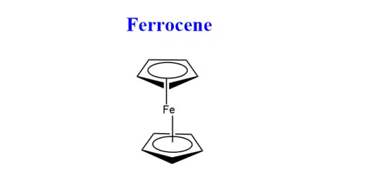
Aka dicyclopentadienyl iron, has the chemical formula (C5H5)2Fe
Ferrocene is the first discovered metallocene sandwich compound.
Metallocene is derived from “Metallo”- transition metal and “Cene” - benzene.
The cyclopentadienyl ligands C5H5 bond in such a way that iron is present in the center of the complex resulting in a sandwich structure
Primarily ferrocene is used as a catalyst in the industrial world
-
Synthesis
Can be prepared: - From Grignard's reagent - From sodium cyclopentadienide - From cyclopentadiene
Grignard's: When the Grignard reagent is treated with iron chloride(II), ferrocene is formed

Sodium Cyclopentadienide: Cyclopentadiene is treated with sodium metal to form cyclopentadienide, which on reacting with FeCl2 forms ferrocene.

Cyclopentadiene: When cyclopentadiene is treated with iron chloride in the presence of a strong base, ferrocene is obtained. Lab method.

-
Physical Properties
Orange crystalline solid at room temp w a camphor like odor.
Stable organometallic compound w melting point 173-174°C and boiling point 249°C.
less soluble in water but soluble in organic solvents like acetone.
-
Chemical Properties
Ferrocene can undergo numerous electrophilic reactions faster than benzene. But, it can not undergo an electrophilic reactions with electrophiles which are strong oxidants like H2SO4 or HNO3.

It can also undergo formylation and carboxylation reactions to give mono-functionalized derivatives, since the functional group extensively deactivates the ferrocenyl group.

Friedel craft acylation reaction can be given by Ferrocene.

--- Solubility Product + Common ion effect
Solubility Product
When a saturated solution of a salt, for example AB, is in contact with the solid phase, the following equilibrium forms:
AgCl (undissolved solid) <--> AgCl (dissolved but unionized) <--> Ag⁺ + Cl⁻ (ions)
AgCl is sparingly soluble in water
The solid is in equilibrium with the ions of solution and this applying the law of mass action, in general: [Ag⁺][Cl⁻]/[AgCl] = K
Now since the solution is saturated and is in contact with the solid, the concentration of the unionized salt molecules should be constant at constant temp.
Hence: [Ag⁺][Cl⁻]/Constant = K where [Ag⁺][Cl⁻] = new constant, [Ag⁺][Cl⁻] = Ksp
Thus, the product of the ionic concentration in a saturated solution of a soluble electrolyte at a constant temp in a constant quantity called solubility product (Ksp)
Ex: using excess reagent, since BaSO4 is insoluble in H2O a small amount would remain in solution if equal amounts of two reagents, like BaCl2 + H2SO4 are taken.
Therefore when carrying out complete precipitation, the precipitant must always be added in a little excess such that the ionic products of compound to be precipitated far exceeds its solubility product.

-
Common Ion Effect
Consider dissociation of a weak electrolyte in water, there exists an equilibrium between ions and unionized molecules to which the law of mass action can be applied.
ex: AB <--> A⁺ + B⁻, [A⁺][B⁻]/[AB] = K (ionization constant)
If the above electrolyte solution added another strong electrolyte with a common ion, either A⁺ or B⁻, the cation or anion will increase.
In order to keep 'K' constant, [AB] should increase in concentration.
This suppression in dissociation is due to a common ion, hence the phenomenon is called common ion effect.
Thus, the suspension of the dissociation of a weak electrolyte in presence of a strong electrolyte which have a common ion with each other is called Common Ion Effect.
-
In group II, cations are ppted as sulphides in acidic medium while in group IV, the cations are ppted as sulphines in basic medium.
The weak electrolyte H2S ionizes in aqueous solution to give sulphide ions in sufficient amounts to exclude the solubility product of group II and the group IV radicals.
But when the solution is made acidic with the addition of dil. HCl, H⁺ ions from the HCl suppresses the ionization of H2S due to the common ion effect
This causes sulphide ion concentration to decrease, thus only the sulphides of group II with a much lower solubility product than the group IV are ppted.
In the presence of NH4OH, the OH- ions combine with the H+ of H2S, forming unionized water, resulting in greater ionization of H2S molecules to give H+ and S-.
This increases the concentration of S- ions, it becomes so high that the solubility product of group IV sulphides is exceeded and this ppted out.
H2S <--> 2H⁺ + S⁻2 is feebly ionized
HCl <--> H⁺ + Cl⁻ is highly ionized
---
Flame test + brown ring test
Flame Test
Flame test is a quick test used in semi-micro analysis to identify the presence of a relatively small number of metal ions in a compound
Flame test is effective in indicating the presence of group I and group V cations
- Principle:
If you excite an atom or an ion by strongly heating, electrons can be promoted from their normal unexcited state into higher orbitals.
As they fall back down to lower levels, energy is released as light.
Each jump involves a specific amount of energy being released as light, and each corresponds to a particular wavelength.
As a result of all these jumps, a spectrum of lines will be produced, some of which will be in the visible part of the spectrum.
The visible color is a combination of all those individual colors.
- Procedure:
To the salt mixture taken in a watch glass, add 2 drops of conc. HCl. Mix with a glass rod to make a paste, expose the end of the rod to the flame of the bunsen burner.
If an apple green color is observed, barium cation from group V may be present.
If a brick red color is observed, calcium cation may be present.
If a crimson red color is observed, strontium cation may be present
-
Brown Ring Test
Brown ring tests is a common nitrate test which determines the presence of nitrate anion in any solution.
- Principle
The nitrate ion functions as an oxidizing agent.
Iron (II) reduces the nitrate ion in the reaction mixture, and iron (II) is oxidized to iron (III).
When nitric oxide is reduced to NO-, ferrous nitroso sulphate complex is formed, which creates a brown ring at the intersection of two layers
- Procedure
A small portion of Na2CO3 extract is taken in a semi-micro test tube
It is acidified by adding a few drops of dil. H2SO4 till no more effervescence is evolved, then followed by a dilute solution of FeSO4.
By holding the test tube at an inclined position, conc. H2SO4 is slowly added along the inner wall of the test tube.
If a brown ring is formed at the junction of the two layers, the presence of nitrate ion is confirmed.

---
Classify organometallic compounds
Organometallic compounds, or organometallics, are compounds where the central metal atom is linked directly with the C atom of the organic ligand.
These compounds contain 1 or more M-C bonds.
The metal atom may be a transition metal, lanthanide, actinide, or a main group element.
Although B, Si, Ge, As, etc. are all non-metallic elements, they are also classified as organometallic compounds.
Cyanides and carbides have M-C bonds, but these compounds are not organometallic compounds because metal is not attached to the carbon of alkyl group.
Classification on the bases of nature of M-C bond: - Ionic bonded - σ-bonded - π-bonded - Bridge bonded
Ionic bonded - The organometallic compounds of highly electropositive metals are usually ionic in nature - In these compounds, the hydrocarbon residue exists as a carbanion with a negative charge, it is attracted to the metal atom by non-directional electrostatic forces. - Colorless, non-volatile, very reactive solids, insoluble in organic solvents. - ex: OMCs with alkali (no lithium), alkaline earth metals, etc. - R2M (M=Ca or Ba), R⁻Na⁺
σ-bonded - C atom of organic ligand bonds to the metal by a 2e⁻, 2 centered covalent bond. - Metals with low electropostive nature form sigma-bonded covalent bonds. - Formed by most elements with values of electropositivity > 1, such as non-metals and weakly electropositive metals. - M and C share a pair of electrons, forming a covalent bond. - ex: (CH3)2Zn
π-bonded - Specific to transition metals - First π-bonded OMC was prepared by ferrocene: (C5H5)2Fe - Ferrocene has a sandwich structure in which the iron atom lies between 2 planar C5H5 rings:

- Bonding involves overlap of π-electrons of the cyclopentadienyl rings with unfilled d-orbitals of the metal.
Bridge-bonded - The compounds in which a loosely bonded electron deficient species exists with the coordination of metals like Li, Be, Al, etc. - This group includes the organometallic compounds with bridging alkyl groups. - Ex: Al2Me6
--- Reactions in HF
Anhydrous HF is a strong acidic solvent
It has high dielectric constant = 84
High dipole moment = 1.9D
Boiling point = 19.4°C and Freezing point = -83°C
It acts as a better solvent than water.
-
Auto Ionization:
Aka self ionization, refers to the process in which a molecules can donate a proton to another molecule of the same species, resulting in the formation of ions.
HF + HF -> H2F⁺ + F⁻
Only a few substances can donate H⁺ to HF:

Precipitation:
Perchlorates, sulphates of non-alkali metals get precipitated when their fluorides dissolved in liq. HF are treated with solution of alkali metal sulphate, perchlorate, etc. NaClO4 + TiF -HF-> TiClO4 ppted + NaF Na2SO4 + NiF2 -HF-> NiSo4 ppted + 2NaF
Acid-Base Reactions:
3HF <--> H2F⁺ (acid) + HF2⁻ (base)
All substances which can form H2F⁺ ion will behave as an acid and substances which yield HF2⁻ ion behave as a base. HNO3(base) + HF -> H2NO3+ + F- BF3(acid) + 2HF -> BF4- + H2F+ HClO4(acid) + HF -> ClO4- + H2F+ HClO4(base) + HF -> H2ClO4+ + F-
Protonation:
Organic compounds like ethanol, benzene, etc. get protonated when dissolved in HF. C2H5OH + HF -> C2H5OH2+ + F- C6H6 + HF -> C6H7+ + H-
---
(5M) Solvolysis reaction of liq. ammonia w ex
Solvolysis is a type of nucleophilic substitution or elimination reaction where the nucleophile is a solvent molecule.
Ammonolysis or ammolysis is solvolysis by ammonia
The attachment of one or more solvent molecules to a solute species through any chemical linkage.
Its products are called solvates H2O -> Hydration -> Hydrates liq. NH3 -> Ammonation -> Ammonites BF3 + NH3 -> BF3.NH3 SiF4 + 2NH3 -> SiF4.2NH3
--- (5M) Bio significance of Na, K, Mg, Cl
Sodium:
Na⁺ cation is a major ion present in the extracellular fluids of organisms, it can be found in large quantities in bones as phosphate. May also be present in chloride and bicarbonate.
Na⁺ ion activates some enzymes in animals.
Imp source for sodium is common table salt (NaCl), which is used in all cooking.
Excessive intake of Na⁺ ions can cause hypertension, for example when fish take in too much saline water where NaCl is in large amounts.
In biological systems, Na⁺ ions regulate acid-base equilibrium.
Sodium is important for the formation of HCl acid in the stomach., the conduction of nerve impulses, and muscle contraction
Since herbivorous animals fail to receive enough Na⁺ ions from their diets, they look out for 'salt-lick' to bridge the gap in their Na⁺ requirements.
Sodium also helps in maintaining osmotic pressure of the body fluid, protecting the organism from fluid loss.
Sodium helps in the prevention of normal irritability of muscle and permeability of cells
Injectable medicines are dissolved in NaCl before they can be administered to humans
Na⁺ cations are essential for both nerve action and heart function.
Na⁺ cation is also responsible for the transport of glucose and amino aids into the cell.
-
Potassium:
K⁺ cation is primarily in intra-cellular fluid as well as extra-cellular fluid.
Imp sources is almost all foods like coffee, tea, cocao, dried beans, molasses, leafy green veg, milk, fish, chicken, pork, dried apricots and peach, banana, orange juice, etc.
K⁺ ion is essential for nerve impulse and muscle contraction.
K⁺ ions are important for all organisms with the potential exception of blue green algae.
When injected intravenously potassium is moderately toxic to mammals.
Much like sodium, potassium also influences acid-base equilibrium in extracellular fluid.
Potassium controls osmotic pressure and water retention
Potassium is essential for metabolic functions such as protein biosynthesis by ribosomes.
Enzymes such as glycolytic enzyme pyruvate kinase requires K⁺ for optimization.
K⁺ ions are required in cells for glucose metabolism, protein synthesis, and activation of some enzymes.
-
Magnesium
Mg is an important non-transition element to all organism. It is present in great concentration in red blood cells.
Imp sources are almonds, cereals, beans, green veg, potatoes, and cheeses.
Mg(II) specifically plays an essential role in many metallo-enzymes
Mg2⁺ cation is present in chlorophyll of plants, enabling the process of photosynthesis.
Constipation, obesity, liver, and gall bladder disorders can all be treated with Mg(II) salts.
Mg forms a vital complex with ATP which is required for most enzymatic reaction involving ATP within the cell.
Magnesium ions are present in important enzymes such as phosphate and aminopeptidase.
Magnesium deficiency is induced by tetanus like cramps and abnormal swelling of the atrial walls in plants. Mg deficiency destroyed the green color of leaves.
-
Chlorine
Cl⁻ ion is an essential electrolyte, commonly found in the body's fluids and blood.
An imp source for sodium is common table salt (NaCl), which is used in all cooking.
Cl⁻ may be present as chlorides (HCl, NaCl, KCl, etc.)
Chlorine is primarily absorbed from the gastrointestinal tract.
Cl⁻ ion helps the body to maintain normal hydration and osmotic pressure, prevents excessive fluid loss.
Cl salts aid in the maintenance of normal acid-base equilibrium.
Chloride ions play an important role in the gaseous transport of CO2.
NaCl and KCl maintain the correct viscosity of blood.
Gastric HCl is an important digestive fluid.
Chlorine ions are mostly excreted by the kidneys in urine.
Deficiency of chlorine in the body leads to hypochloremia: poor appetite, increased urination, irritability, muscle twitching
Excess of chlorine in the body leads to hyperchloremia: diarrhea, vomiting, fatigue, dehydration
--- (5M) Reactions for identification of CO3(2-) and NH4+ ions
Analysis of NH4⁺ cation:
Ammonium salts when titrated with NaOH solution produces ammonia gas with a pungent odor.
NH4Cl + NaOH -> NaCl + NH3 + H2O
The evolution of NH3 gas allows us to confirm that NH4⁺ is present.
To confirm the evolved gas in NH3:
i) Expose the gas to a filter paper paper dipped in CuSO4 solution - If the paper turns blue, the gas in NH3 and HN4⁺ is initially confirmed. - CuSO4 + 4NH3 -> [Cu(NH3)4]SO4 - [Cu(HN3)4]SO4 is tetramine copper complex, it is a deep blue color.
ii) Expose the gas to a filter paper dipped in Nessler's reagent. - If the paper turns brown, NH4⁺ is confirmed. - Ammonium chloride reacts with Nessler's reagent to form a brown ppt called iodide of Millon's base.

-
Analysis of CO3(2-) anion
Carbonate salts when treated with dilute HCl acid produces carbon dioxide gas along with brisk effervescence.
Na2CO3 + 2HCl -> 2NaCl + CO2 +H2O
Confirming that the gas emitted is CO2 will confirm the presence of CO3(2-).
If we pass the gas through lime water and the lime water turns milky, the gas if confirmed to be CO2 and CO3(2-) is confirmed.
CO2 + Ca(OH)2 (lime water) -> CaCO3 + H2O
---
Chlorophyll Structure


---
Grignard's Reagent


0 notes
Text
Pharmaceutical RO + EDI Water Treatment Systems: Ensuring Ultra-Pure Water for Critical Applications
Pharmaceutical industries require water of exceptional purity for various processes, including drug formulation, cleaning, and production. One of the most effective methods to achieve this high level of water purity is through Pharmaceutical RO (Reverse Osmosis) + EDI (Electrodeionization) Water Treatment Systems. These systems are integral in producing Purified Water (PW) and Water for Injection (WFI), which are critical in ensuring that pharmaceutical products meet stringent quality and safety standards.
Understanding Reverse Osmosis in Pharmaceutical Water Treatment
Reverse Osmosis (RO) is a well-established technology for removing a wide range of impurities from water, including dissolved salts, organics, and particulates. In the pharmaceutical industry, RO is commonly used as the primary treatment step due to its ability to remove over 99% of contaminants, ensuring that only the purest water is available for further processing.
In an RO system, water is forced through a semi-permeable membrane that allows water molecules to pass while blocking larger molecules and impurities. This process effectively removes:
Dissolved salts and ions
Microorganisms
Organic compounds
Endotoxins
The result is high-purity water that serves as the feedwater for the next stage: Electrodeionization (EDI).
The Role of Electrodeionization (EDI) in Water Treatment
Electrodeionization (EDI) is a water purification technology that further polishes the water after the RO stage. EDI operates by using electrical current and ion-exchange resins to remove residual ions from the RO-treated water. Unlike conventional ion-exchange methods, EDI continuously regenerates its resins without the need for chemical additives, making it a more sustainable and cost-effective solution.
EDI systems are highly effective in reducing ionic contaminants to extremely low levels, often meeting the USP (United States Pharmacopeia) and EP (European Pharmacopeia) standards for pharmaceutical-grade water. Some of the contaminants targeted by EDI include:
Cations such as calcium, magnesium, and sodium
Anions like chloride, sulfate, and nitrate
Weakly ionized substances, such as silica and carbon dioxide
Through this combined approach of RO + EDI, the resulting water meets the strict regulatory requirements for pharmaceutical manufacturing.
Benefits of RO + EDI Systems for Pharmaceutical Applications
The combination of RO and EDI technologies in pharmaceutical water treatment systems offers several key benefits, making them indispensable in pharmaceutical production:
Consistent Water Quality: RO + EDI systems ensure a reliable supply of ultra-pure water, essential for sensitive pharmaceutical processes such as sterile drug formulation and preparation of injectable solutions.
Compliance with Regulatory Standards: The water produced by these systems meets the stringent quality standards set by regulatory bodies such as the FDA, USP, and EP. This compliance ensures the safety and efficacy of pharmaceutical products.
Chemical-Free Operation: EDI eliminates the need for chemical regeneration of ion-exchange resins, reducing operational costs and environmental impact.
Reduced Risk of Contamination: RO + EDI systems minimize the presence of microorganisms and endotoxins, reducing the risk of contamination in water used for pharmaceutical production.
Scalability and Flexibility: These systems can be customized to meet the specific needs of different pharmaceutical processes, from small-scale laboratory applications to large-scale manufacturing.

Key Components of a Pharmaceutical RO + EDI Water Treatment System
The effectiveness of Pharmaceutical RO + EDI Water Treatment Systems relies on several key components that work together to ensure the highest level of water purity:
Pre-treatment Systems: Before water enters the RO system, it typically undergoes pre-treatment to remove larger particles, chlorine, and other impurities that could damage the RO membranes. Common pre-treatment methods include multimedia filtration, activated carbon filtration, and softening.
RO System: The RO unit is the heart of the system, responsible for removing the majority of dissolved solids and impurities. Modern pharmaceutical RO systems are designed with advanced features such as high-rejection membranes and energy recovery devices to maximize efficiency.
EDI System: After RO treatment, the water is passed through the EDI unit, where ion-exchange resins and electrical current work together to remove any remaining ionic impurities.
Storage and Distribution Systems: Purified water from the RO + EDI system is typically stored in stainless steel or high-density polyethylene (HDPE) tanks to prevent recontamination. A high-purity water distribution system ensures that the purified water is delivered to various points of use within the pharmaceutical facility.
Monitoring and Control Systems: Modern RO + EDI systems are equipped with sophisticated monitoring and control systems that continuously track water quality parameters such as conductivity, pH, and temperature. This ensures that any deviations in water quality are detected and corrected in real time.
Maintenance and Validation of RO + EDI Systems
Maintaining the performance of RO + EDI systems is crucial in ensuring the consistent production of high-purity water. Pharmaceutical companies must adhere to strict maintenance schedules, including routine inspections, cleaning of RO membranes, and regular testing of water quality.
In addition, validation is a critical aspect of pharmaceutical water systems. Regulatory authorities require that all water systems used in drug manufacturing be validated to ensure they consistently produce water that meets quality standards. This involves rigorous testing and documentation, including performance qualification, operational qualification, and installation qualification of the water system.
Applications of RO + EDI Systems in the Pharmaceutical Industry
Pharmaceutical RO + EDI Water Treatment Systems play a vital role in various pharmaceutical processes, including:
Sterile Water for Injection (WFI): RO + EDI systems are essential in producing WFI, which is used for the preparation of injectable medications. WFI must meet stringent microbial and endotoxin limits to ensure the safety of patients.
Cleaning and Sterilization: High-purity water is required for cleaning pharmaceutical equipment, ensuring that no contaminants are introduced during the production process. RO + EDI systems provide water of the necessary quality for equipment cleaning and sterilization.
Oral and Topical Drug Formulations: In the production of non-injectable drugs, purified water is used as an ingredient in the formulation of oral and topical medications. RO + EDI systems ensure that the water used in these formulations is free from impurities that could compromise product quality.
Biotechnology and Biopharmaceuticals: Biopharmaceutical production requires ultra-pure water for cell culture, fermentation, and protein purification processes. RO + EDI systems provide the water quality necessary to support these sensitive processes.
Environmental Impact and Sustainability
Pharmaceutical manufacturers are increasingly focused on sustainability, and RO + EDI water treatment systems offer several environmental benefits. The elimination of chemical regenerants in EDI systems reduces the environmental footprint associated with traditional ion-exchange methods. Furthermore, modern RO systems are designed with energy-efficient technologies, reducing the overall energy consumption of the water treatment process.
In addition to their environmental benefits, RO + EDI systems contribute to overall cost savings by reducing the need for consumables such as chemicals and replacement filters.
Pharmaceutical RO + EDI Water Treatment Systems are essential for producing the ultra-pure water required in pharmaceutical manufacturing. These systems ensure that water used in drug formulation, equipment cleaning, and other critical processes meets the stringent purity standards mandated by regulatory authorities. By combining the strengths of Reverse Osmosis and Electrodeionization, pharmaceutical companies can achieve consistent, high-quality water production while reducing their environmental impact.
For pharmaceutical companies seeking reliable and compliant water treatment solutions, SWJAL PROCESS Pvt. Ltd. stands as a leading Pharmaceutical RO + EDI Water Treatment Systems manufacturer in Mumbai, India, providing expertise and advanced technologies tailored to the specific needs of the pharmaceutical industry.
#pharmacutical industry#water treatment system#RO + EDI water treatment Plant#manufacturer#SWJAL PROCESS#India
0 notes
Text
Weather Control Technology: The Future of Climate Management

The idea of controlling the weather using technology has been transformed from what was once a fantasy into a real scientific field. Even though the application of weather manipulation on a large scale is still a theoretical construct, research is being done into promising technologies and methods with encouraging results. As global climate change and natural disasters are on the rise, weather modification technologies can be the key for easing these issues.
1. Cloud Seeding
Cloud seeding is the most beloved weather control technique and also the most commonly known one. To that end, substances like silver iodide or salt are spread off into clouds to including rain. In several countries, cloud seeding has been used for drought mitigation, water supply to increase, and fog control at airports. But, doubts are still there regarding its long-run effectiveness and environmental impacts.
2. Hurricane Modification
Studies are being carried out on ways to alter the direction of hurricanes or weaken them. Research is being done on such techniques as injecting cold water into the ocean or using wind turbines to disrupt hurricane winds. These are hypothetical methods, and as such, they may be able to stop storms from causing mass destruction, though it is hard to tell.
3. Laser Technology
Laser-based weather control is a novel branch of science, that can allow humans to decide the course of lightning storms in the future. Scientists are studying the process of using high-energy lasers to ionize the air which creates a path of desirable lightning to land safe on the ground. This technology has the potential to save people from lightning strikes which is the major problem of severe storms.
4. Geoengineering
Geoengineering is a larger set of technologies intended to manipulate the Earth’s climate to balance the consequences of global warming. The idea of solar radiation management is one way to do this; where particles are released into the stratosphere to reflect sunlight and cool the planet. The use of this method may act as a shield to global warming but most definitely, the long-term ecological impacts of the interventions are still largely unknown.
Ethical Considerations and Challenges
Unintentional consequences
For instance, weather modification may hurt the normal weather patterns, which will result in rainfall shifts in the neighboring areas, or it may disturb the natural ecosystems.
Impact on the environment
The impending development of geoengineering projects introduces threats to the environment, such as loss of biodiversity and the alteration of the ocean currents.
Ethical dilemmas
The capability to manipulate the weather brings about grave ethical concerns regarding the just distribution of the benefits and the potential for abuse.
Benefits
Food security will arise as a result of weather control as it acts to enhance the production of crops.
Preventing disaster: Employing these strategies ensures that the consequences of climate-related disasters are greatly diminished.
Economic Development: Protection of the buildings and the materials.
Challenges
Environmental Impact: Unanticipated outcomes of weather control.
Ethical Concerns: The question of who has the right to control the weather.
Technical Limitations: Limitations in the technology for scalability and efficacy.
Emerging Trends
Space-Based Weather Control: Solar radiation management through the use of satellites.
Artificial Weather Systems: The grand scheme of weather modification.
Nanotechnology: Weather alteration at the molecular scale.
AI-Powered Weather Forecasting: The ability to predict with greater accuracy.
The Future of Weather Control
Even though weather control technology is still in the early stages, it has some potential for alleviating serious global issues such as climate change and food security. Nevertheless, moving ahead with caution and reflecting on the ethical as well as environmental consequences of such actions are of utmost importance.
Conclusion
Weather control technology sits on the borderline of science and ethical dilemmas. Although the promise of tackling global challenges is attractive, it is necessary to treat weather control technology with caution and respect the ecological and ethical aspects. As science and technology progress, there must be an open discussion about responsible weather modification practices.
#weathercontrol#climateengineering#technology#science#innovation#technews#controversialtopics#ethicalissues#debate#discussion#futuretechnology#futurism
0 notes
Text
How Does an MRI Scan Work: A Comprehensive Guide for First-Time Patients
Magnetic Resonance Imaging (MRI) is a powerful diagnostic tool that uses magnetic fields and radio waves to create detailed images of the body's internal structures. If you’re preparing for your first MRI scan, it’s natural to feel a little anxious or uncertain about the process. Understanding how an MRI works, what to expect during the procedure, and how it can help in diagnosis can ease your concerns. This guide provides a comprehensive overview of MRI, including a step-by-step explanation, preparation tips, and answers to common questions.
What is an MRI Scan?
An MRI scan is a non-invasive medical imaging technique that helps doctors diagnose a variety of conditions, from injuries to diseases affecting the brain, spinal cord, joints, and other soft tissues. Unlike X-rays or CT scans, MRI does not use ionizing radiation. Instead, it employs a strong magnetic field and radiofrequency pulses to generate highly detailed images.
MRI is particularly effective in imaging soft tissues, which makes it ideal for diagnosing issues in organs like the brain, heart, liver, and muscles.
How Does an MRI Work?
1. Magnetic Field Generation
The MRI scanner consists of a large cylindrical magnet. When you lie inside the scanner, the magnetic field aligns the hydrogen atoms in your body. Since the human body is largely composed of water, and water contains hydrogen atoms, this alignment occurs throughout your entire body.
2. Radiofrequency Pulses
Once the hydrogen atoms are aligned, the MRI machine sends radiofrequency pulses to disturb this alignment. When the pulses are turned off, the hydrogen atoms realign to the magnetic field, releasing energy as they do so. The MRI machine detects this energy and uses it to create images of your body’s internal structures.
3. Image Formation
The data from the energy released by hydrogen atoms is processed by a computer to generate detailed cross-sectional images of your organs, tissues, and bones. The images are incredibly precise, allowing radiologists to identify abnormalities or monitor the progress of conditions.
What to Expect During Your MRI Scan
1. Preparation
Clothing: You’ll be asked to change into a hospital gown and remove any metal objects, including jewelry, watches, and hairpins, as these can interfere with the magnetic field.
Contrast Agents: In some cases, a contrast agent (usually gadolinium) may be injected into your bloodstream to enhance the visibility of certain structures or abnormalities in your body.
Communication: You will be able to communicate with the MRI technician during the scan via an intercom system.
2. The Procedure
Positioning: You will lie down on a motorized bed that slides into the MRI scanner. It’s crucial to remain still throughout the procedure to ensure clear images.
Duration: Depending on the part of the body being scanned, an MRI can last anywhere from 20 minutes to over an hour.
Noise: MRI machines make loud knocking or tapping noises due to the switching of magnetic fields. You’ll be given earplugs or headphones to protect your ears from the noise.
Sensation: The procedure is completely painless. Some patients may feel slight warmth in the area being scanned, but this is normal.
3. After the Scan
Once the scan is complete, you can resume normal activities immediately. If a contrast agent was used, drinking extra water can help flush it out of your system more quickly. Your doctor will review the MRI images and discuss the results with you.
Common Uses of MRI
MRI scans are used to diagnose or monitor various medical conditions, including:
Neurological Disorders: MRI is the gold standard for imaging the brain and spinal cord. It helps in diagnosing conditions like multiple sclerosis, brain tumors, and spinal injuries.
Musculoskeletal Injuries: Soft tissue injuries, such as ligament tears or cartilage damage, are best visualized with MRI. It’s commonly used in sports medicine and orthopedic assessments.
Cardiac Conditions: Cardiovascular MRI provides detailed images of the heart, helping to detect heart disease, inflammation, or abnormalities in the heart's structure.
Cancer: MRI is often used in cancer diagnosis and monitoring, particularly for breast, liver, and prostate cancers.
MRI Safety Considerations
While MRI scans are generally safe, there are a few safety considerations to keep in mind:
Metal Implants: If you have metal implants like pacemakers, cochlear implants, or certain types of metal plates, let your doctor know, as the magnetic field could interfere with these devices.
Pregnancy: MRI is usually avoided in the first trimester of pregnancy unless absolutely necessary.
Claustrophobia: Some patients may feel claustrophobic inside the narrow MRI machine. If you’re anxious, speak with your doctor about options such as an open MRI or mild sedation.
Factors That Can Influence MRI Scan Costs
When planning for an MRI, it’s also important to be aware of the factors that can influence the cost of the scan. For example, the type of MRI (with or without contrast), the location of the facility, and the complexity of the scan can all impact the final price. To learn more about the key factors that contribute to MRI scan costs, you can refer to this comprehensive guide from Clinico Pathology Lab & Diagnostic Centre: Factors Influencing MRI Scan Cost: What to Consider.
This article explains the various elements that affect the pricing of an MRI, including:
Type of MRI Machine: Advanced MRI machines with high-field magnets or specialized features may increase the cost.
Use of Contrast: MRI scans that require contrast agents are generally more expensive than those without.
Location of the Facility: Prices can vary significantly based on whether you are getting an MRI at a private clinic, public hospital, or specialized diagnostic center.
Additional Services: Some facilities may include extra services like fast-track appointments, radiologist consultations, or follow-up scans, which can contribute to higher costs.
By understanding these factors, you can make informed decisions and better plan for any diagnostic imaging needs.
Benefits of MRI Scans
One of the primary benefits of MRI is its ability to provide detailed images without exposing patients to radiation. This makes it especially valuable for imaging delicate or sensitive areas like the brain, spinal cord, and joints. MRI can also detect abnormalities that other imaging techniques, such as X-rays or CT scans, might miss, allowing for earlier diagnosis and more effective treatment planning.
Conclusion
If you’re a first-time patient scheduled for an MRI scan, understanding the process can help alleviate any anxiety you might have. MRI is a safe, non-invasive, and highly effective diagnostic tool used to identify and monitor a wide range of medical conditions. By preparing for your scan and knowing what to expect, you can approach the procedure with confidence.
If you are concerned about the cost of an MRI scan or would like more detailed information about factors that can influence pricing, don't forget to check out Clinico’s in-depth guide on Factors Influencing MRI Scan Cost. This resource provides valuable insights to help you navigate the financial aspects of your diagnostic journey.
0 notes
Text
Understanding Magnetic Resonance Imaging (MRI): A Guide to the Powerful Diagnostic Tool
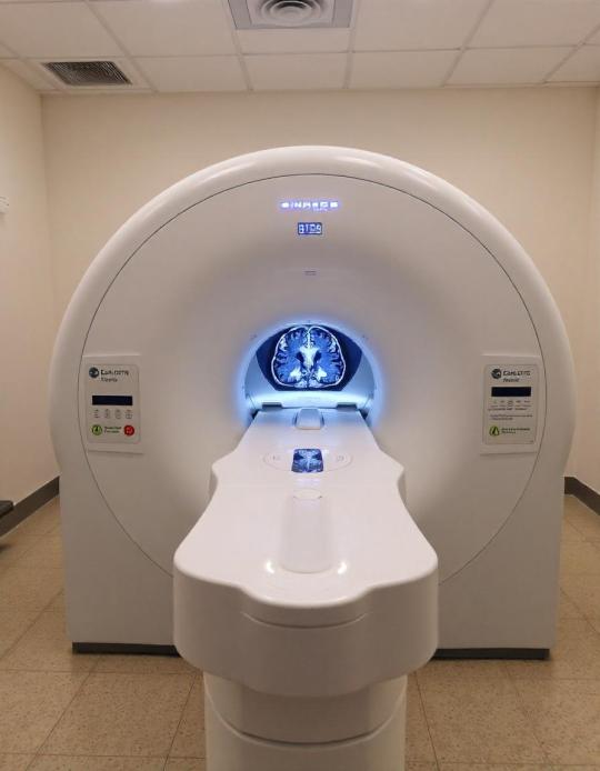
Magnetic Resonance Imaging (MRI) is one of the most advanced and widely used imaging technologies in modern medicine. It plays a crucial role in diagnosing a variety of medical conditions, allowing healthcare providers to view detailed images of the inside of the body without the need for invasive procedures. Whether it's for detecting abnormalities in the brain, spine, joints, or internal organs, MRI provides a clear and precise picture, guiding physicians in making informed decisions.
How MRI Works
MRI uses strong magnetic fields and radio waves to generate detailed images of organs, tissues, and structures within the body. Unlike X-rays or CT scans, which use ionizing radiation, MRI relies on the magnetic properties of atoms, particularly hydrogen, which is abundant in the body's water and fat molecules. The process involves aligning the hydrogen atoms using a powerful magnet, then disrupting that alignment with radio waves. As the atoms return to their normal state, they emit signals that are captured and converted into highly detailed images by the MRI machine.
Key Uses of MRI
MRI is an incredibly versatile diagnostic tool and can be used for a wide range of medical applications, including:
Brain and Nervous System: MRI is often used to diagnose conditions such as brain tumors, strokes, aneurysms, multiple sclerosis, and spinal cord injuries.
Musculoskeletal System: For injuries or conditions involving muscles, ligaments, and joints, MRI provides a clear view of soft tissues, helping in the diagnosis of torn ligaments, cartilage damage, and bone infections.
Cardiovascular System: MRI can provide detailed images of the heart and blood vessels, assisting in the diagnosis of heart disease, damaged heart tissue, or issues with blood flow.
Abdominal and Pelvic Organs: MRI is commonly used to examine organs such as the liver, kidneys, pancreas, and reproductive organs, identifying issues like tumors, infections, or abnormalities.
Benefits of MRI
Non-invasive: MRI is a painless and non-invasive procedure, making it a safe option for patients.
Detailed Images: MRI offers superior image clarity, especially for soft tissues, providing more detailed information than other imaging techniques.
No Radiation Exposure: MRI does not use harmful ionizing radiation, making it a safer option for repeated use, especially for sensitive populations like children or pregnant women.
Preparing for an MRI
While MRI is a safe procedure, there are a few important considerations:
Metal Objects: Because of the strong magnetic field, patients must remove all metal objects before the scan. Those with metal implants, pacemakers, or certain medical devices may need to consult their doctor before undergoing an MRI.
Claustrophobia: Some patients may feel claustrophobic in the enclosed MRI machine. Open MRI scanners are available in some facilities, and doctors can provide sedation if needed.
Contrast Agents: In some cases, a contrast agent may be injected to enhance the visibility of certain tissues. While generally safe, patients with kidney issues or allergies should inform their healthcare provider beforehand.
Conclusion
Magnetic Resonance Imaging (MRI) اشعة رنين مغناطيسي is a vital tool in modern healthcare, offering detailed and accurate images to aid in the diagnosis and treatment of a wide range of medical conditions. Its non-invasive nature and lack of radiation make it a preferred option for many physicians and patients alike. With advancements in MRI technology continuing to evolve, this diagnostic tool will remain at the forefront of medical imaging, providing critical insights into patient health.
0 notes
Text


First results from 2021 rocket launch shed light on aurora’s birth
Newly published results from a 2021 experiment led by a University of Alaska Fairbanks scientist have begun to reveal the particle-level processes that create the type of auroras that dance rapidly across the sky.
The Kinetic-scale Energy and momentum Transport experiment — KiNET-X — lifted off from NASA’s Wallops Flight Facility in Virginia on May 16, 2021, in the final minutes of the final night of the nine-day launch window.
UAF professor Peter Delamere’s analysis of the experiment’s results was published Nov. 19 in Physics of Plasmas.
“The dazzling lights are extremely complicated,” Delamere said. “There’s a lot happening in there, and there’s a lot happening in the Earth’s space environment that gives rise to what we observe.
“Understanding causality in the system is extremely difficult, because we don’t know exactly what’s happening in space that’s giving rise to the light that we observe in the aurora,” he said. “KiNET-X was a highly successful experiment that will reveal more of the aurora’s secrets.”
Want more? Read the dramatic story of the KiNET-X mission in 12 short installments that include videos, animations and additional photographs.
One of NASA’s largest sounding rockets soared over the Atlantic Ocean into the ionosphere and released two canisters of barium thermite. The canisters were then detonated, one at about 249 miles high and one 90 seconds later on the downward trajectory at about 186 miles, near Bermuda. The resulting clouds were monitored on the ground at Bermuda and by a NASA research aircraft.
The experiment aimed to replicate, on a minute scale, an environment in which the low energy of the solar wind becomes the high energy that creates the rapidly moving and shimmering curtains known as the discrete aurora. Through KiNET-X, Delamere and colleagues on the experiment are closer to understanding how electrons are accelerated.
“We generated energized electrons,” Delamere said. “We just didn’t generate enough of them to make an aurora, but the fundamental physics associated with electron energization was present in the experiment.”
The experiment aimed to create an Alfvén wave, a type of wave that exists in magnetized plasmas such as those found in the sun’s outer atmosphere, Earth’s magnetosphere and elsewhere in the solar system. Plasmas — a form of matter composed largely of charged particles — also can be created in laboratories and experiments such as KiNET-X.
Alfvén waves originate when disturbances in plasma affect the magnetic field. Plasma disturbances can be caused in a variety of ways, such as through the sudden injection of particles from solar flares or the interaction of two plasmas with different densities.
KiNET-X created an Alfvén wave by disturbing the ambient plasma with the injection of barium into the far upper atmosphere.
Sunlight converted the barium into an ionized plasma. The two plasma clouds interacted, creating the Alfvén wave.
That Alfvén wave instantly created electric field lines parallel to the planet’s magnetic field lines. And, as theorized, that electric field significantly accelerated the electrons on the magnetic field lines.
“It showed that the barium plasma cloud coupled with, and transferred energy and momentum to, the ambient plasma for a brief moment,” Delamere said.
The transfer manifested as a small beam of accelerated barium electrons heading toward Earth along the magnetic field line. The beam is visible only in the experiment’s magnetic field line data.
“That’s analogous to an auroral beam of electrons,” Delamere said.
He calls it the experiment’s “golden data point.”
Analysis of the beam, visible only as a varying shades of green, blue and yellow pixels in Delamere’s data imagery, can help scientists learn what is happening to the particles to create the dancing northern lights.
The results so far show a successful project, one that can even allow more information to be gleaned from its predecessor experiments.
“It’s a question of trying to piece together the whole picture using all of the data products and numerical simulations,” Delamere said.
Three UAF students doing their doctoral research at the UAF Geophysical Institute also participated. Matthew Blandin supported optical operations at Wallops Flight Facility, Kylee Branning operated cameras on a NASA Gulfstream III aircraft out of Langley Research Center, also in Virginia, and Nathan Barnes assisted with computer modeling in Fairbanks..
The experiment also included researchers and equipment from Dartmouth College, the University of New Hampshire and Clemson University.
youtube
3 notes
·
View notes
Text
Is an ultrasound procedure safe?

Here’s why:
Non-invasive: Ultrasound imaging is non-invasive, meaning it doesn’t involve the use of needles, injections, or surgical incisions. Instead, it relies on high-frequency sound waves to produce images of internal structures
No Radiation: Unlike X-rays and CT scans, ultrasound imaging doesn’t expose patients to ionizing radiation, reducing the risk of radiation-related side effects or long-term health concerns.
Painless: Ultrasound procedures are typically painless and well-tolerated by patients. The procedure involves applying a gel to the skin and moving a transducer over the area of interest to capture images, causing no discomfort.
Real-time Imaging: Ultrasound provides real-time imaging, allowing healthcare providers to observe internal structures and organs as they function, aiding in the diagnosis of various conditions such as pregnancy, vascular diseases, and abdominal issues.
Versatility: Ultrasound imaging can be used to examine various parts of the body, including the abdomen, pelvis, heart, blood vessels, and musculoskeletal system, making it a versatile tool in healthcare.
Safe for Pregnant Women: Ultrasound is commonly used during pregnancy to monitor fetal development and assess the health of the mother and baby. Extensive research has shown that ultrasound imaging is safe for both the pregnant woman and the developing fetus when performed by trained professionals.
Minimal Side Effects: The risks associated with ultrasound procedures are minimal. In rare cases, patients may experience mild discomfort from the pressure of the transducer on the skin or slight heating of the tissues during prolonged examinations. However, these effects are temporary and pose no significant harm.
Diagnostic Accuracy: Ultrasound imaging offers high diagnostic accuracy in many clinical scenarios, aiding in the detection and diagnosis of various conditions without the need for invasive procedures or exposure to radiation.
Overall, ultrasound procedures are considered safe and effective diagnostic tools, playing a crucial role in modern healthcare for both diagnostic and therapeutic purposes. However, as with any medical procedure, it’s essential to have ultrasound examinations performed by qualified healthcare professionals to ensure optimal safety and accuracy.
0 notes
Text
Brain MRI Scan in Delhi NCR
Magnetic Resonance Imaging (MRI) of the brain is one of the most advanced and non-invasive diagnostic tools used in modern medicine. It provides highly detailed images of the brain’s internal structures, allowing doctors to detect abnormalities, diagnose neurological conditions, and plan treatments with precision.
Unlike X-rays or CT scans that use radiation, brain MRI uses a powerful magnetic field and radio waves to produce high-resolution images of the brain. It is safe, painless, and widely used to investigate a variety of symptoms, including headaches, seizures, dizziness, memory loss, and more.
What Is a Brain MRI?
A brain MRI is a specialized scan that captures detailed images of the brain and surrounding tissues. It is often recommended when a patient presents with unexplained symptoms related to the brain or nervous system. During the scan, the patient lies on a table that slides into a large, tunnel-shaped machine. The MRI machine creates a strong magnetic field that aligns hydrogen atoms in the body. Then, radio waves are used to disturb this alignment. As the atoms return to their normal position, they emit signals that are captured to form detailed images of the brain.
Why Is a Brain MRI Done?
Brain MRI is commonly used to:
Investigate chronic headaches or migraines
Evaluate unexplained neurological symptoms like dizziness, vision problems, or weakness
Diagnose brain tumors or cysts
Detect strokes or bleeding in the brain
Monitor multiple sclerosis (MS) or other degenerative brain diseases
Examine brain infections like encephalitis or abscesses
Assess brain injuries after trauma
Diagnose epilepsy and identify the source of seizures
Check brain development in children
Unlike CT scans, which may miss small or subtle lesions, MRI offers greater detail, especially for soft tissue structures like the brain and spinal cord.
Types of Brain MRI
Depending on the specific medical need, different types of MRI sequences and techniques can be used, including:
Contrast MRI: A contrast dye (gadolinium) is injected to enhance the visibility of blood vessels, tumors, and inflammation.
Functional MRI (fMRI): Measures brain activity by detecting changes in blood flow. Useful in mapping brain functions before surgery.
Magnetic Resonance Angiography (MRA): Focuses on blood vessels in the brain to detect aneurysms or blockages.
Diffusion Tensor Imaging (DTI): Maps nerve fiber pathways in the brain, helpful in assessing traumatic brain injuries.
Benefits of Brain MRI
Non-Invasive and Painless: The scan does not require surgery or incisions. It is completely painless and typically takes 30 to 60 minutes.
Radiation-Free: Unlike X-rays or CT scans, MRI does not use ionizing radiation, making it safer for repeated use, especially in children and pregnant women (after the first trimester).
Highly Detailed Images: MRI provides clearer and more detailed images of the brain compared to other imaging techniques. This is crucial for detecting small abnormalities.
Early Diagnosis of Serious Conditions: Brain MRI can detect life-threatening conditions like tumors, aneurysms, or strokes at an early stage, improving treatment outcomes.
Helps in Treatment Planning: From identifying surgical targets to monitoring the progression of neurological diseases, MRI plays a critical role in planning and evaluating treatment effectiveness.
Preparation and What to Expect
Before the scan, patients are typically asked to remove any metallic objects such as jewelry, glasses, or hearing aids. Those with metal implants, pacemakers, or clips may not be eligible for an MRI, as the magnetic field can interfere with these devices.
The patient lies still inside the MRI scanner while the machine creates a series of thumping or tapping noises. Earplugs or headphones are often provided to reduce discomfort from the noise. In some cases, a contrast dye may be injected to enhance image clarity.
It’s important to stay still during the scan to avoid blurry images. Claustrophobic patients may be offered sedation or an open MRI, depending on availability.
Risks and Limitations
Brain MRI is extremely safe, but there are a few considerations:
Claustrophobia: Some patients may feel uncomfortable in the confined space.
Metal Implants: Patients with certain implants or devices may not be suitable candidates.
Allergic Reaction to Contrast: Rarely, patients may react to the contrast dye, although it’s usually mild and manageable.
Despite these minor concerns, the benefits of brain MRI far outweigh the risks in most cases.
A brain MRI is a powerful diagnostic tool that allows doctors to look inside the brain with remarkable clarity. Whether it’s detecting a tumor, identifying the source of a seizure, or evaluating a stroke, MRI provides critical information that can save lives and improve treatment outcomes. Its safety, accuracy, and non-invasive nature make it an essential part of modern neurology and diagnostic imaging.
To know the MRI scan cost in Delhi NCR visit Healthi India
0 notes
Text
Everything You Need to Know About MRI - Alsafwa Radiology Center
Introduction
Magnetic Resonance Imaging, commonly known as MRI, is a powerful diagnostic tool that provides detailed images of the body's internal structures. This non-invasive procedure has revolutionized the medical field by allowing physicians to diagnose and monitor a wide range of conditions with remarkable precision. At Alsafwa Radiology Center, we pride ourselves on offering state-of-the-art MRI services, ensuring our patients receive the highest standard of care. In this comprehensive guide, we will explore everything you need to know about MRI scans, including their purpose, process, benefits, and the advanced technology available at our center.
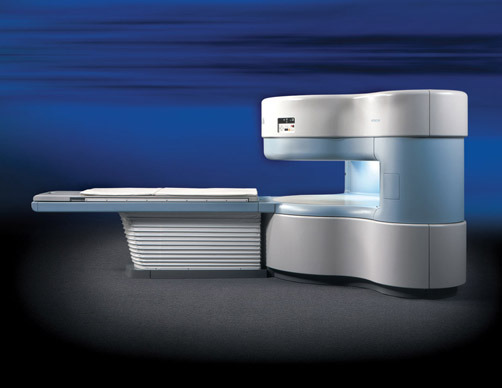
What is an MRI Scan?
An MRI scan is a medical imaging technique that uses a magnetic field and radio waves to create detailed images of the organs and tissues within the body. Unlike X-rays and CT scans, which use ionizing radiation, MRI scans are radiation-free, making them a safer option for many patients.
How Does an MRI Scan Work?
MRI technology relies on the principles of nuclear magnetic resonance. Here’s a step-by-step breakdown of how an MRI scan works:
Magnetic Field Creation: The MRI machine generates a strong magnetic field, which aligns the hydrogen atoms in the body.
Radio Wave Pulses: The machine then emits pulses of radio waves, which knock the hydrogen atoms out of their aligned position.
Signal Detection: When the radio waves are turned off, the hydrogen atoms return to their original alignment, releasing energy in the process.
Image Formation: This released energy is detected by the MRI sensors and used to create detailed images of the internal structures.
Uses of MRI Scans
MRI scans are versatile and can be used to diagnose and monitor a variety of conditions, including:
Brain and Spinal Cord Issues: MRI is crucial for detecting brain tumors, multiple sclerosis, stroke, and spinal cord injuries.
Joint and Musculoskeletal Problems: It helps in diagnosing torn ligaments, cartilage issues, and other musculoskeletal conditions.
Abdominal and Pelvic Conditions: MRI scans can identify liver disease, kidney problems, and reproductive system disorders.
Cardiovascular Concerns: MRI can evaluate the heart and blood vessels, helping in the diagnosis of congenital heart defects, aneurysms, and other cardiovascular conditions.
Benefits of MRI Scans
Non-Invasive and Painless: MRI scans are non-invasive, meaning they do not require any incisions or injections (unless contrast is used), and they are generally painless.
No Radiation Exposure: Unlike X-rays and CT scans, MRI scans do not use ionizing radiation, making them a safer alternative for repeated imaging, especially in children and pregnant women.
High-Resolution Images: MRI provides highly detailed images, which can lead to more accurate diagnoses and better treatment planning.
Types of MRI Machines
At Alsafwa Radiology Center, we offer various types of MRI machines to cater to different patient needs:
Traditional MRI: The conventional MRI machine features a long, narrow tube. While highly effective, it can be uncomfortable for patients with claustrophobia or those with larger body sizes.
Open MRI: Our Open MRI machines provide a more comfortable and less claustrophobic experience, making them ideal for children, elderly patients, and those with anxiety.
Functional MRI (fMRI): fMRI is used to measure and map brain activity by detecting changes in blood flow. It is particularly useful in neuroscience research and pre-surgical planning.
Preparing for an MRI Scan
Preparing for an MRI scan involves a few simple steps to ensure the procedure goes smoothly:
Consultation: Your physician will provide detailed instructions and address any concerns you may have before the scan.
Clothing and Jewelry: You will be asked to remove any metal objects, such as jewelry and belts, as metal can interfere with the magnetic field.
Contrast Agents: In some cases, a contrast agent may be administered to enhance the visibility of certain tissues or blood vessels. This is typically done through an intravenous injection.
Communication: It’s important to inform the technician if you have any implants, pacemakers, or metal fragments in your body, as these can pose risks during the scan.
The MRI Scan Process
Here’s what you can expect during an MRI scan at Alsafwa Radiology Center:
Positioning: You will lie down on a movable table that slides into the MRI machine. Our technicians will ensure you are comfortable and provide pillows or cushions if needed.
Scanning: The machine will produce a series of loud noises as it takes images. You will be given earplugs or headphones to reduce the noise.
Stillness: It’s crucial to remain as still as possible during the scan to ensure clear images. Our technicians will communicate with you throughout the process to provide instructions and support.
Duration: An MRI scan typically lasts between 30 to 60 minutes, depending on the area being examined.
After the MRI Scan
Once the scan is complete, you can resume your normal activities immediately. The images will be reviewed by a radiologist, who will then share the results with your physician. Your doctor will discuss the findings with you and determine the next steps in your care plan.
Why Choose Alsafwa Radiology Center for Your MRI Scan?
At Alsafwa Radiology Center, we are committed to providing exceptional care and the latest in medical imaging technology. Here’s why you should choose us for your MRI scan:
State-of-the-Art Technology: We use advanced MRI machines that deliver high-quality images with minimal discomfort to patients.
Expert Staff: Our team of radiologists and technicians are highly trained and experienced, ensuring accurate results and a smooth experience.
Patient-Centered Care: We prioritize patient comfort and convenience, offering flexible scheduling and a supportive environment.
Comprehensive Services: In addition to MRI, we offer a wide range of imaging services, including X-rays, CT scans, and ultrasound, making us a one-stop center for all your diagnostic needs.
Conclusion
MRI scans are a critical tool in modern medicine, providing detailed and accurate images that aid in the diagnosis and treatment of numerous conditions. At Alsafwa Radiology Center, we are dedicated to offering the highest standard of MRI services, with a focus on patient comfort and cutting-edge technology. Whether you need an MRI for a routine check-up or a specific medical concern, you can trust our team to provide exceptional care every step of the way.
For more information or to schedule an appointment, contact Alsafwa Radiology CenterAlsafwa Radiology Center today. Let us help you take the next step in your healthcare journey with confidence and peace of mind.
0 notes
Text
How HPLC Column Works: Principles of Separation Explained
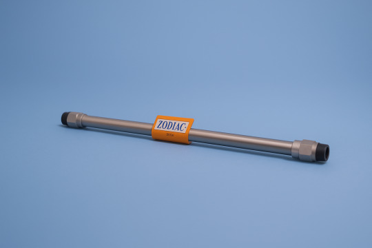
Introduction
High-Performance Liquid Chromatography (HPLC) is a cornerstone technique in analytical chemistry, widely used in pharmaceutical, biotech, food, and environmental labs. At the heart of every HPLC system lies a critical component: the column. But how does an HPLC column actually work? Understanding this process helps analysts improve separation, troubleshoot issues, and choose the right column for their applications.
In this article, we’ll break down how an HPLC column works, covering its internal structure, separation mechanism, and key factors that affect performance.
The Role of the HPLC Column
The HPLC column is the site where the actual separation of analytes occurs. It contains a packed bed of stationary phase particles that interact with compounds in the injected sample. As the mobile phase flows through the column, different compounds move at different speeds, depending on how strongly they interact with the stationary phase.
The result? Each component elutes at a different time, producing a unique retention time that’s essential for identification and quantification.
How HPLC Column Works – Step-by-Step Process
1. Sample Injection
The process starts when the sample is introduced into the HPLC system via an injector and carried by the mobile phase toward the column.
2. Interaction with the Stationary Phase
As the sample passes through the column, each analyte interacts with the stationary phase. The strength and nature of these interactions (hydrophobic, polar, ionic) determine how long a compound is retained.
3. Separation Mechanism
In Reverse Phase HPLC (e.g., C18 columns), non-polar compounds interact more strongly with the hydrophobic stationary phase and elute later.
In Normal Phase HPLC, polar compounds are retained longer.
4. Elution
Each compound exits (elutes from) the column at a different time. The detector records these elution times as peaks on a chromatogram.
Internal Structure of an HPLC Column
An HPLC column consists of:
Tube material: Usually stainless steel or PEEK
Stationary phase: Silica-based particles bonded with functional groups (e.g., C18, C8, phenyl, amino, etc.)
Particle size: Typically 3–5 µm in analytical columns; affects resolution and backpressure
Column dimensions: Common lengths (50–250 mm) and internal diameters (2.1–4.6 mm)
Each of these factors influences how well the column separates your target analytes.
Factors That Influence Column Functionality
Several variables impact how effectively an HPLC column performs: Factor Impact
Mobile Phase Composition Affects elution strength and retention time
pH Alters ionization of analytes and stability of bonded phase
Temperature Influences viscosity, analyte interaction, and column lifetime
Flow Rate Affects resolution and analysis time
Optimizing these conditions is crucial for method development and reproducible results.
Common Questions: How HPLC Columns Work
❓ Is a longer column always better?
Not necessarily. Longer columns may improve resolution but increase run time and pressure. It's about balancing separation needs with practicality.
❓ Can I reuse the same column for different samples?
Columns can be reused, but cross-contamination and degradation over time can impact results. Using guard columns helps extend column life.
❓ What’s the difference between analytical and preparative columns?
Analytical columns are used for small-scale quantification, while preparative columns are larger and used to isolate and collect purified compounds.
Conclusion
Understanding how an HPLC column works is fundamental to mastering liquid chromatography. From the chemistry of interactions to the mechanical design, each element of the column plays a role in achieving sharp, reliable separation.
At Zodiac Life Sciences, we offer a wide range of HPLC columns — including C18, C8, phenyl, and amino — engineered for precision, consistency, and longevity. Explore our selection today to find the right column for your lab's needs.
0 notes